Which of the Following Indicates Possible Ms During an Mri
MRI and MS. Higher risk of developing the disease.
Brain Sciences Free Full Text Pattern Recognition Of The Multiple Sclerosis Syndrome Html
When patients get an MRI they will typically be given gadolinium contrast.

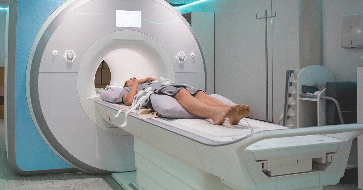
. Magnetic resonance imaging MRI is the diagnostic tool that currently offers the most sensitive non-invasive way of imaging the brain spinal cord or other areas of the body. One would expect however that the abnormalities would show up in the MRI eventually in a patient with MS MRI Mimickers of MS. Relapsing-remitting MS RRMS.
Magnetic resonance imaging MRI was formally included in the diagnostic work-up of patients presenting with a clinically isolated syndrome CIS suggestive of multiple sclerosis MS in 2001 by an International Panel of experts. In an attempt to assure the highest sensitivity and specificity a set of guidelines referred to as the McDonald criteria 1 utilizes magnetic resonance imaging MRI to provide supportive data to facilitate the diagnosis of MS. However if there is inflammation that reduces the BBB the contrast is able to get into the brain and will show up as actively enhancing on inflammed lesions.
Its thought to be the result of an immune system attack. Examining the results from an MRI a spinal tap blood tests and evoked potentials measurements of electrical activity in certain areas of the brain and spinal cord against the McDonald criteria help doctors discern between MS and other diseases that can. 1 MS diagnosis requires demonstration of disease dissemination in space DIS and time DIT and exclusion of other conditions that can mimic.
Two or more relapses plus evidence of damage in at least two CNS regions. A formal diagnosis of MS can be made without any. RRMS is the most common type and it involves flares during which symptoms get worse and times of remission when they go away almost completely before returning.
Blood tests are used to rule out other autoimmune diseases and disorders or. MS causes demyelination or the damage of myelin. One of the best aspects of an MRI is that its non-invasive other than the administration of the gadolinium gd into a vein.
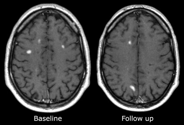
Which type of ms is the most common type of the disease relapsing. Magnetic resonance imaging MRI plays a crucial role in multiple sclerosis MS diagnosis disease monitoring prognostication and research. The MRI can also be used to confirm that damage has occurred at two different points in time.
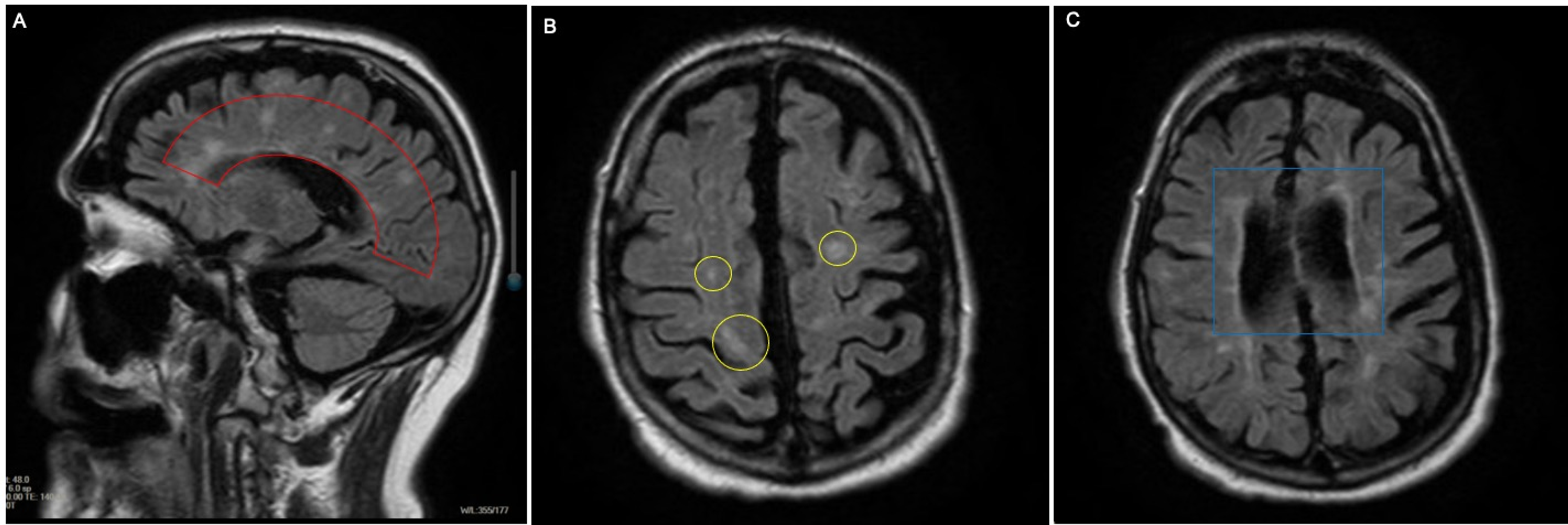
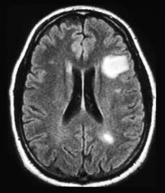
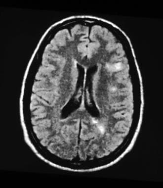
1 Alternatively there may be white matter lesions that might be seen. When what of the substance nigra cells stop producing dopamine Parkinsons disease motor symptoms begins to accur. MRI scans can identify lesions that occur due to MS.
The MRI by itself cant determine if you have MS since some other medical conditions could cause spots to show up during procedure. As scar tissue lesions. Though the vast majority of MS patients have abnormalities on brain MRI an estimated 5 of patients have normal imaging.
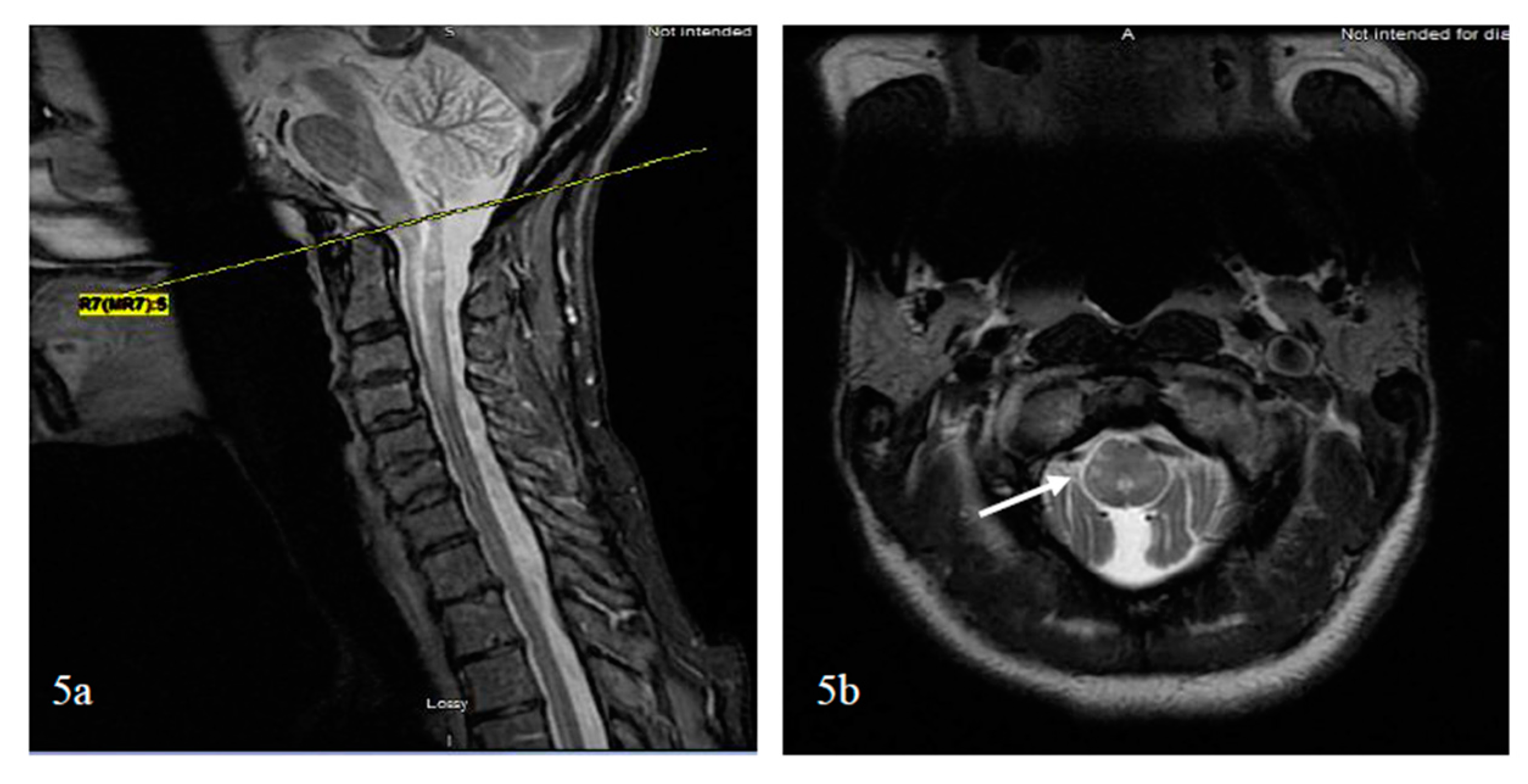
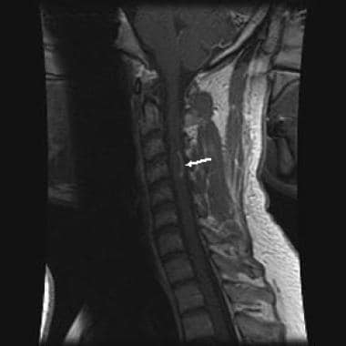
In one study 35-mm axial T2-weighted images with full spinal cord coverage showed 22 more lesions in patients with MS than 3-mm sagittal scans especially for lesions with small axial diameters. The MRI can be used to look for a second area of damage in a person who has experienced only one attack also called a relapse or an exacerbation of MS-like symptoms referred to as clinically-isolated syndrome CIS. MRI has been part of the International Panel criteria for the diagnosis of MS since 2001 and its use has become increasingly vital as.
Diagnosing MS can be difficult because there is no definitive way to tell if a person has it. There are also conditions that can show up on a brain MRI and look like multiple sclerosis. Scans can let healthcare professionals know when.
MRI has made it possible to visualize and. 39 Post-mortem studies have shown unambiguously that MTR is strongly associated with the percentage of. In short the MRI isnt necessarily the be-all end-all diagnostic tool for determining the presence of multiple.
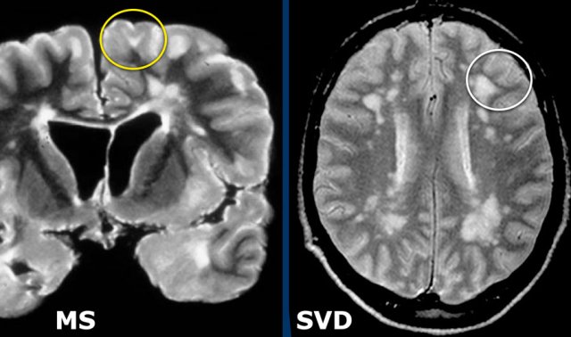
The MS Lesion Checklist differs from Barkhof criteria for MS Box in 2 key aspects. In the case of your husband they may signify small vessel ischemic disease we also refer to this as white matter diseasebasically the small vessels in the brain are showing signs of ischemia. Multiple sclerosis MS is a condition in which the bodys immune system attacks the protective covering myelin surrounding the nerves of the central nervous system CNS.
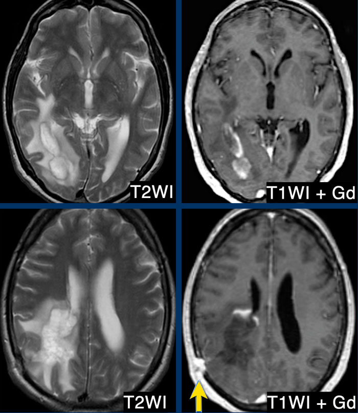
As I have stated in my post not all white matter lesions on the MRI scan indicate multiple sclerosis. Both criteria for dissemination in time and space have been fulfilled. Several important practice guidelines updates for MRI in MS have been published recently including the 2017 revised McDonalds Criteria1 Magnetic Resonance Imaging in MS network guidelines2 and revised recommendations of.
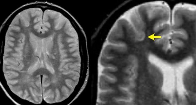
8 Barkhof criteria were not designed to be applied to patients without suspicion of MS. It is the preferred imaging method to help establish a diagnosis of MS and to monitor the course of the disease. MS lesions can show white matter inflammation demyelination and scarring or sclerosis.
Magnetisation transfer MRI measures the interactions between protons in free fluids and protons bound to macromolecules by use of the magnetisation transfer ratio MTR whereby a low MTR is an indicator of damage to myelin and axonal membranes. 7 First Barkhof imaging criteria were created to predict development of MS in a patient with clinically isolated syndrome CIS that suggest inflammatory demyelination a clinical syndrome typical of MS. The sensitivity of T 1 - and T 2-weighted MRI to white matter WM lesions and to regional as well as global atrophy has made this modality central to the diagnosis and treatment monitoring of multiple sclerosis MS1-4 Paradoxically these findings correlate only weakly with clinical disability an incongruity due MRIs insensitivity to microscopic pathology and lack of.
This is the protective layer that surrounds your nerve fibers. What treatment category of MS focuses on reducing the symptoms of MS. Normally this contrast stays within the blood and does not get into the brain because of the BBB.
The Radiology Assistant Multiple Sclerosis 2 0
Cureus Complications Of Covid 19 Pneumonia And Multiple Sclerosis Exacerbation

Multiple Sclerosis Ms Types Symptoms And Causes

Paediatric Multiple Sclerosis And Antibody Associated Demyelination Clinical Imaging And Biological Considerations For Diagnosis And Care The Lancet Neurology

Magnetic Resonance Imaging Mri Ms Trust
Ms Brain Lesions Pictures Symptoms And More

Ms Lesions On The Spine Connection Diagnosis And Treatment

Value Of 3t Susceptibility Weighted Imaging In The Diagnosis Of Multiple Sclerosis American Journal Of Neuroradiology
Multiple Sclerosis Ms Diagnosis What Tests Are There
The Radiology Assistant Multiple Sclerosis 2 0
Multiple Sclerosis Clinical Presentation History Physical Examination Clinical Rating Scales
Which Findings On Mri Suggest Multiple Sclerosis Ms
The Radiology Assistant Multiple Sclerosis 2 0

Repair Of Multiple Sclerosis Brain Damage May Be Possible
Multiple Sclerosis Clinical Presentation History Physical Examination Clinical Rating Scales

White Matter Imaging An Overview Sciencedirect Topics

Multiple Sclerosis Tests And Diagnosis
The Radiology Assistant Multiple Sclerosis 2 0

Mri Scans Definition Uses And Procedure


Comments
Post a Comment